Introduction
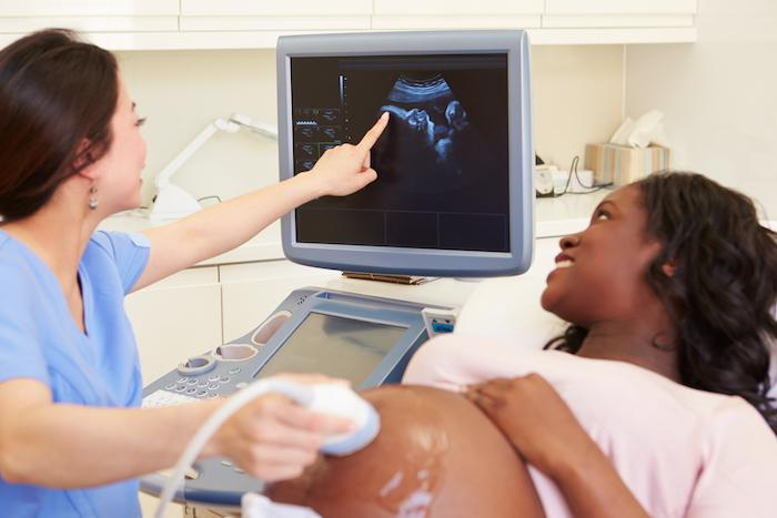
In the realm of medical diagnostics, few technologies have captured the imagination and awe-inspiring beauty like ultrasound imaging. From its humble beginnings to the cutting-edge techniques used today, ultrasound has revolutionized the field of medical imaging, providing a window into the human body like never before. In order to ensure the well-being of their developing baby, the couple decided to undergo an early pregnancy scan essex, seeking reassurance and early detection of any potential concerns. In this article, we will explore the captivating images revealed by pioneering ultrasound techniques, showcasing the remarkable advancements and the impact they have had on healthcare.
The Birth of Ultrasound Imaging
Ultrasound imaging, also known as sonography, emerged as a medical diagnostic tool in the mid-20th century. Inspired by the echolocation abilities of dolphins and other marine mammals, scientists and researchers sought to harness the power of sound waves for medical purposes. The realization that sound waves could penetrate the human body and bounce back to create detailed images marked the birth of ultrasound imaging.
Early Ultrasound Devices: The Quest for Clarity
The first ultrasound devices were crude compared to the sophisticated technology we have today. Early pioneers used simple transducers and piezoelectric crystals to emit and receive sound waves. These devices provided basic images with limited resolution and detail, primarily used in obstetrics to monitor fetal development.
Advancements in Transducer Technology
Over the years, significant advancements in transducer technology have propelled ultrasound imaging to new heights. The development of phased-array and linear-array transducers revolutionized the field, enhancing image quality and expanding the scope of diagnostic applications.
Phased-array transducers, consisting of multiple elements, allowed for beamforming—a technique that focuses sound waves on specific areas of interest. This innovation improved image resolution, providing clearer and more detailed visualizations. Linear-array transducers, on the other hand, offered high-resolution imaging capabilities, enabling the examination of intricate anatomical structures.
From 2D to 3D: The Depth of Detail
One of the most captivating advancements in ultrasound imaging is the transition from two-dimensional (2D) to three-dimensional (3D) imaging. Traditional 2D ultrasound captures images in slices, providing a flat representation of structures. However, the introduction of 3D imaging added depth and realism to the images, enhancing the diagnostic capabilities.
By acquiring a series of 2D images from different angles, sophisticated algorithms reconstruct a 3D representation of the scanned area. This innovation has proven particularly valuable in obstetrics, allowing parents to see detailed renderings of their unborn babies and fostering a deeper connection with the growing life within.
The Wonders of 4D Ultrasound
Building upon the foundation of 3D imaging, the advent of 4D ultrasound introduced the element of time into the equation. This groundbreaking technique captures real-time 3D images, showcasing the dynamic movements and interactions of organs and structures within the body.
Expectant parents are now able to witness their baby’s movements in real-time, from tiny kicks and stretches to facial expressions. The ability to observe the developing fetus with such clarity and realism has created profound emotional connections and enhanced the prenatal bonding experience.
Specialized Ultrasound Techniques: Beyond the Surface
Ultrasound imaging has extended its reach beyond routine examinations, leading to specialized techniques tailored for specific medical fields. Some notable advancements include:
1. Doppler Ultrasound
Doppler ultrasound, a technique based on the Doppler effect, enables the visualization and analysis of blood flow within the body. By evaluating the velocity and direction of blood flow, doctors can identify blockages, assess vascular conditions, and detect abnormalities in organs and tissues. Doppler ultrasound has become an essential tool in cardiovascular diagnostics and plays a crucial role in monitoring fetal well-being during pregnancy.
2. Contrast-Enhanced Ultrasound (CEUS)
Contrast-enhanced ultrasound involves the use of contrast agents to improve the visibility of blood vessels and enhance diagnostic accuracy. By injecting microbubble-based contrast agents into the bloodstream, ultrasound images become more vivid and detailed. CEUS has found applications in various fields, including liver imaging, tumor characterization, and assessing vascular abnormalities.
3. Elastography
Elastography is a technique that evaluates tissue stiffness, aiding in the diagnosis of diseases affecting organ elasticity. By applying external pressure or vibration and measuring the resulting tissue deformation, elastography provides valuable insights into conditions such as liver fibrosis, breast lesions, and prostate abnormalities. This technique has the potential to reduce the need for invasive procedures and improve diagnostic accuracy.
Beyond Diagnostic Imaging: Ultrasound-Guided Procedures
Ultrasound imaging not only serves as a diagnostic tool but also guides a range of minimally invasive procedures. Using real-time imaging, doctors can perform precise needle insertions, biopsies, and aspirations with improved accuracy and safety.
Ultrasound-guided procedures have become standard in various medical specialties, including interventional radiology, anesthesia, and pain management. The ability to visualize internal structures and precisely target treatment areas has transformed the landscape of minimally invasive interventions.
Conclusion: Unveiling the Marvels of Ultrasound Imaging
From its humble beginnings to the sophisticated techniques of today, ultrasound imaging has transcended the boundaries of traditional diagnostics. Through the use of advanced transducers, 3D and 4D imaging, and specialized techniques like Doppler ultrasound and elastography, ultrasound has revealed captivating images that have revolutionized medical practice.
These pioneering ultrasound techniques not only provide valuable diagnostic information but also offer a window into the marvels of the human body. They foster deeper connections between patients and their healthcare providers and empower individuals to actively participate in their own healthcare journeys.
As we continue to unlock the potential of ultrasound imaging, the future holds even more promise. Advancements in artificial intelligence, miniaturization of devices, and integration with other imaging modalities will further enhance the accuracy, accessibility, and scope of ultrasound in healthcare.
The captivating images revealed by pioneering ultrasound techniques showcase the miracles hidden within our bodies, reminding us of the power of innovation and the wonders of modern medicine.